
You are here: Home / Case study - CT scan of chest (P + C)
CT Scan of chest (P + C)
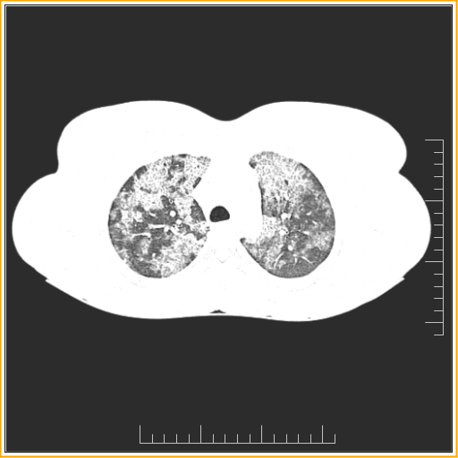
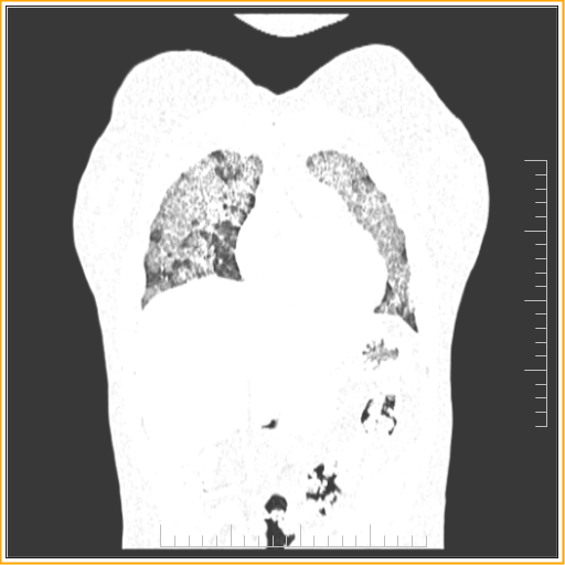
PATIENT: 23 year old female
COMPLAINT: AKT taken before 2 month, cough, fever, chest pain, hemoptosis and weight loss
Patchy areas of ground glass opacities with smooth inter and intra lobular septal thickening (crazy paving pattern) diffusely distributed throughout of both lungs with small areas of intervening normal lung parenchyma at places predominantly in upper lobe.
Finding of suggestive of interstitial lung disease likely pulmonary alveolar proteinosis